 “I have been doing pathology for last many years, and I still love it. I am interested in learning about non-invasive diagnostic imaging techniques of skin like confocal microscopy” said Dr. Maria Shevchuk, MD, FCAP, an Associate Professor of Pathology and Laboratory Medicine at Weill Cornell Medical College and Associate Attending Pathologist at New York Presbyterian Hospital. On Wednesday, May 25 Dr. Maria visited NIDIskin Rao Dermatology New york to learn about skin confocal microscopy.
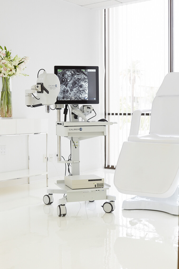
During the meeting Dr. Babar K. Rao explained the importance of confocal microscopy in 21st century and why he prefers to do confocal microscopy before biopsy especially to rule out malignant cutaneous tumours. Dr. Baba K. Rao is a board certified Dermatologist, Dermatopathologist, and Mohs Surgeon, and is a leading authority on pigmented lesions, as well as a pioneer in Dermoscopy and Confocal Microscopy.
“Whenever we do a biopsy, obviously it is much happier for everyone to give the patient a benign diagnosis, but on the other hand, this also means that whatever has been removed did not need to be removed, and the patient unnecessarily endured the discomfort of the biopsy and the subsequent anxiety of waiting for a diagnosis.” told Dr. Rao.
Dr. Rao then said “ So my ultimate goal is to perform an invasive procedure only when its is necessary meaning, start with dermoscopy, confocal microscopy or any another tool you may have to save the patient from anxiety, pain and a scar. Statistically, we already perform far less biopsies in our offices than any other office in the region. Even more importantly, Non-Invasive Diagnostic Imaging of the Skin allows us to analyse skin lesions quickly and painlessly, and may help to catch worrisome lesions that would otherwise go undetected.”
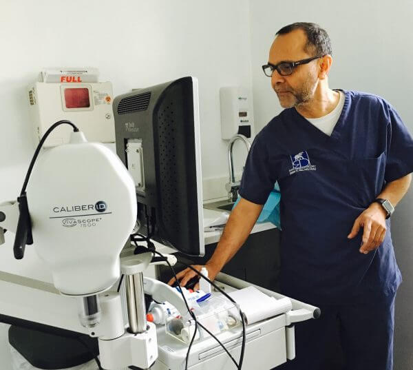 During her visit Dr. Maria learned the techniques to perform skin confocal microscopy and how Dr. Rao read the confocal images . “The most important question in all confocal meetings until now was how long does it take to perform confocal microscopy. Now I know it’s a very simple procedure.” said Dr. Maria. She then saw how Dr. Rao completed his confocal diagnosis reports. “This is my virtual tray just like histology slides tray in pathology.” said Dr. Rao while signing his confocal cases through vivanet work list.
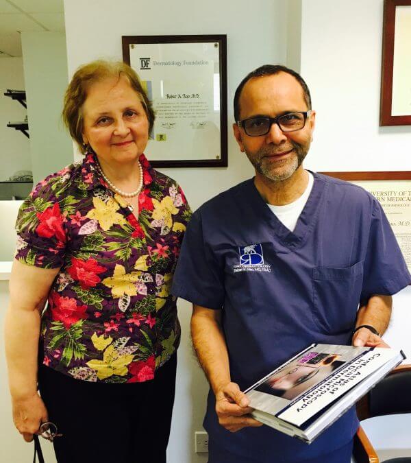 At the end of the visit Dr. Rao presented “Atlas of Confocal Microscopy in Dermatology by Dr. Babar K. Rao” to Dr. Maria. “I am very excited that Dr. Rao has published atlas of confocal microscopy.” said Dr. Maria.