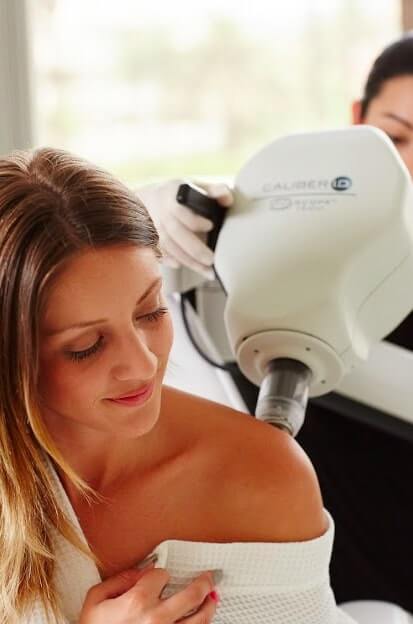
 Reflectance confocal microscopy (RCM) is a non-invasive and painless imaging technique that enables in vivo visualization of cellular details of skin. The resolution of this innovative technique is almost comparable to traditional histology. Unlike biopsy RCM enables the investigation of entire lesion.
How Does RCM Work?
Reflectance confocal microscope is a FDA 510(k) Cleared Class I Laser Device. This device uses a diode laser as a monochromatic light. No adverse events have been reported in 500 clinical studies.
Why we do RCM?
Until recently, whenever a mole or spot was suspected malignant, a surgical biopsy was required to rule out the skin cancer. Although this has been the standard protocol for skin cancer detection and prevention, the vast majority of skin biopsies of moles in the United States result in benign diagnosis, which means that surgery- and any discomfort and scarring associated with it-was unnecessary. RCM can eliminate most of these unnecessary biopsies.
What Exactly is the RCM Procedure?
It is as simple as 1, 2, and 3. At NIDIskin, we systematically rule out benign moles as candidates for biopsy using a 3-step process.
Step 1: Investigation of all moles and mark suspicious moles or spots.
Step 2: Capture magnified and illuminated images of any moles that require monitoring with VivaCam™ Dermatoscopy.
Step 3: Examine cellular structure of any high-risk moles with VivaScan™ Confocal Microscopy
No biopsy is required for the mole or spot diagnosed as benign on RCM by our experts.
Talk to the Experts
If you are concerned about any mole or spot on your body, talk with the professionals at NIDIskin about ways to reduce your chances of biopsy. For more information call us at 212-949-0393. For weekly updates like our facebook page at https://www.facebook.com/nidiskin.
